How tech is transforming the way we treat stroke patients. Atul Gupta, MD, Chief Medical Officer and Interventional Radiologist, Philips Image Guided Therapy, discusses the latest innovations in diagnosing strokes
By The Engine Room, Monday 15 July 2019

Stroke affects someone on earth every two seconds and has little respect for age (one-third of stroke victims in the US are younger than 65). Stroke’s ischemic aftermath leaves behind a path of devastation, not just to patients but also to their families. Alongside a high mortality rate, those who survive a stroke all too often experience life-changing and debilitating disability. The economic impact of stroke is vast, costing an estimated $34 billion each year in the US alone. Fortunately, significant new clinical data, coupled with notable advances in medical technology, have ushered in an exciting period of change, one that may well revolutionise stroke practice. Time has long been of the essence when it comes to treating stroke – the typical patient loses almost two million neurons each minute in which stroke is untreated, so time is indeed brain. Physicians responding to a patient with suspected stroke will be all too familiar with the ‘golden window’, the period of time in which patients can have a thrombectomy, where the tiny clot capable of such massive destruction is removed through a minimally-invasive image-guided therapy (IGT) procedure. By removing the clot, the flow of blood to the brain is restored, potentially reversing or substantially reducing the long-term effects of stroke.
More time available
This golden window has long been deemed to be just six hours. This limited timeframe meant that many patients who could and should have been treated sadly were not. However, research in May 2018 indicated the potential to extend this treatment window by up to four times. The landmark DEFUSE 3 and DAWN trials showed that thrombectomy could be performed effectively in selected patients up to 24 hours after a stroke. While the trial findings are being met with significant excitement from physicians and medical societies, it also means that a larger pool of patients will be eligible for the procedure. Yet few hospitals are equipped with either the appropriate technology or interventional physicians to meet the anticipated demand. It is further indication that we need new ways to ensure that patients are correctly diagnosed, with access to the right expertise, facilities and after-care, within the restricted timeframe. This was further reflected in a recent policy statement published by the American Stroke Association which demands improvements in stroke systems of care to reflect the advances in scientific knowledge and innovations in clinical care. It’s a powerful call to ensure the significant developments that have been made to facilitate optimal stroke care delivery and improve patient outcomes. Notably, one of the recommendations in the policy statement called for emergency workers to carefully consider which hospital can give stroke patients the best care, within a fifteen-minute journey, highlighting the importance of optimum treatment in this time-sensitive situation. There’s no doubt that there is great progress being made. The ‘stroke ambulance’ – an ambulance equipped with computed tomography (CT) imaging technology to clarify whether the patient has had a stroke or not and whether they are a candidate for thrombectomy. If deemed a suitable candidate, the patient can be swiftly diverted to the nearest stroke centre capable of performing a thrombectomy. Already in use in several cities, including New York and Berlin, the stroke ambulance clearly saves time, but it is neither cost-efficient or practical to equip all ambulances with such capabilities.
New technology
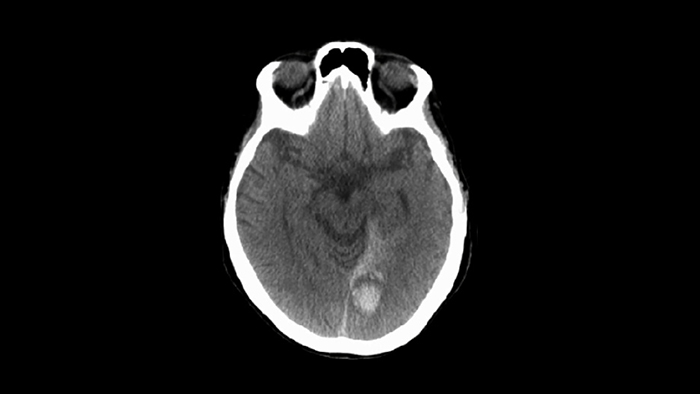
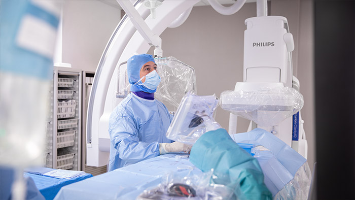
In some cases, hospitals have combined a dedicated CT scanner with an angiography system into the interventional suite, creating a ‘one-stop stroke shop’ for patients suspected of having a stroke. Allowing patients to be routed from the ambulance directly to the interventional suite reduces time but it’s neither scalable nor a practical solution, given the high costs involved. What could be a realistic and cost-effective solution? Already, some so-called one-stop stroke shop interventional suites are availing of advanced cone beam CT – an imaging tool that is integrated into interventional systems such as Philips’ Azurion. This advanced technology allows for the creation of a high-quality CT image of the brain, without the need for an actual CT scanner. Data presented at the 2018 Society of NeuroInterventional Surgery (SNIS) proved that Philips’ cone beam CT compares favourably with conventional CT scans and decrease delays in care by up to 60 minutes, important at a time when every moment can make a difference.
An intriguing alternative that’s currently in development is what could be described as a portable ‘stroke visor’. Based on non-invasive bioimpedance spectroscopy, these diagnostic devices have shown up to 92 per cent accuracy in the diagnosis of large vessel occlusion, the type of stroke which responds to thrombectomy. They can be carried by emergency medical service workers and placed onto patients’ heads while in transit. Early data is certainly promising, but more data is needed before the devices are commercially available.
Faster care
While it’s clear that we are making strides with the technological approaches to increasing our chances of improving outcomes for stroke patients, it’s clear that what we also need are more trained experts who can implement both these innovative solutions and the infrastructure in place across the stroke continuum. A smart example of this can be seen in a forward-thinking stroke centre in the Hague where a dynamic team of neurologists, interventionalists, diagnostic radiologists, and more, are cross-collaborating and using innovations like our Azurion image-guided therapy system to expedite the treatment of stroke patients, reducing stroke-to-needle time and improving patient care. The ongoing fight against stroke is one that needs to be tackled on multiple fronts. It’s encouraging to see the progress that’s been made so far. An exciting future in the field of stroke research lies ahead.
Looking ahead, imaging and interventions relating to stroke will remain high on the agenda. Equally, ongoing testing and development of solutions will bring us closer to making the very most of the golden window. In a bid to speed up acute care for stroke patients, Karolinska University Hospital, together with Philips, is developing a communication tool that allows specialists to exchange patient information even quicker. The goal is to ensure that everyone involved has the right information to hand to help ensure the patient has access to the fastest and best possible care.